Wrist Fracture Case File
Wrist Fracture Case File
Eugene C. Toy, MD, Lawrence M. Ross, MD, PhD, Han Zhang, MD, Cristo Papasakelariou, MD, FACOG
CASE 3
A 23-year-old male reports that during a game of hoops (basketball), he tripped while driving the ball to the basket, and fell on his outstretched right hand with the palm down. Two days later, he phoned his anatomist father and related that his right wrist was painful. Later that day, he visited his father, who noted that the wrist was slightly swollen and tender but without deformity. He instructed his son to extend the right thumb, thereby accentuating the anatomical “snuffbox,” which is extremely tender to deep palpation. His father advised him to get his hand and wrist x-rayed.
⯈ What is the most likely diagnosis?
⯈ What is the most likely anatomic defect?
ANSWERS TO CASE 3:
Wrist Fracture
Summary: A 23-year-old male trips while playing basketball and suffers trauma to the right wrist. The wrist is slightly swollen, tender, but not deformed. However, deep palpation of the anatomical snuffbox elicits extreme tenderness.
• Most likely diagnosis: Wrist fracture
• Most likely anatomical defect: Fracture of the narrow middle portion of the scaphoid carpal bone
CLINICAL CORRELATION
This young man tripped while playing basketball and stretched out his right hand to protect himself. His hand, with the palm down and probably deviated to the side of the radius, took the brunt of the fall, resulting in significant impact force to the wrist. This resulted in pain and swelling of the wrist, especially on the radial side, with point tenderness deep in the anatomical snuffbox. This is the common mechanism for a fracture of the scaphoid carpal bone, the most commonly fractured carpal bone. Point tenderness over a bone or bony process is a hallmark of a fracture at that site. Radiologic confirmation of a fracture is important. The scaphoid bone has a unique blood supply, and proper reduction and alignment of the segments is necessary to decrease the risk of avascular necrosis. A fall on an outstretched hand such that it produces hyperextension of the wrist may result in dislocation of the lunate bone. The lunate is usually displaced anteriorly into the carpal tunnel and may impinge on the median nerve. The lunate is the most commonly dislocated carpal bone. A fall on an outstretched palm may also result in a transverse fracture of the distal radius or a Colles fracture, which produces a dorsal displacement of the distal fragment, resulting in the characteristic “dinner fork” (also termed “bayonet”) deformity. A Smith fracture of the radius in the same region of younger individuals is less common. In a Smith fracture, there is trauma to the dorsal aspect of a flexed wrist, and the wrist is deformed with the distal radial fragment displaced ventrally in a “spade” deformity.
APPROACH TO:
The Wrist
OBJECTIVES
1. Be able to describe the bones and joints of the wrist
2. Be able to describe the anatomy of the radius and ulnar as it relates to the transmission of forces in the upper limb and its effect on the forearm bones
3. Be able to describe the boundaries of the anatomical snuff box and its clinical significance
DEFINITIONS
ANATOMICAL SNUFFBOX: Depression on the lateral aspect of the wrist formed by the tendons of the extensor pollicis brevis and abductor pollicis longus anteriorly and the extensor pollicis longus posteriorly
FRACTURE: A break in the normal integrity of a bone or cartilage
AVASCULAR NECROSIS: Death of cells, tissues, or an organ due to insufficient blood supply
DISCUSSION
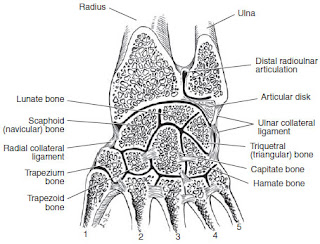
The junction of the forearm and hand, called the wrist region, is a complex of several joints. The articulation of the distal radius with the ulna, called the distal radioulnar joint, is the site of movement of the radius anteriorly around the ulna during pronation. The radius and ulna are united by an articular disk or triangular fibrocartilage and associated ligaments, which intervenes between the ulna and carpal bones. The wrist joint proper is formed between the distal radius, the triangular fibrocartilage, and the proximal row of carpal bones. The eight carpal bones are arranged in proximal and distal rows of four bones each. From lateral to medial, the proximal row is composed of the scaphoid, lunate, triquetrum, pisiform, and the distal row is composed of the trapezium, trapezoid, capitate, hamate (mnemonic: “Some ladies try perfume that they can’t handle”). Approximately 50 percent of movement at the wrist occurs at the wrist joint proper, with the remaining 50 percent occurring at the intercarpal joint, between the two rows of carpal bones. A capsule, reinforced by palmar and dorsal radiocarpal ligaments, surrounds the joint. The radial collateral ligament strengthens the capsule laterally and limits adduction (ulnar deviation). The ulnar collateral ligament strengthens the capsule medially and limits abduction (radial deviation) (Figure 3-1).
In addition to the distal radioulnar joint, the proximal radioulnar joint allows pivotal movement of the radius with the humerus and the ulna during pronation and supination. The radius and ulna are also joined by the interosseous membrane and its fibers to form a syndesmosis. The individual fibers are attached proximally on the radius but distally on the ulna. Impact forces on an outstretched hand are transmitted at the wrist to the radius, through the interosseous membrane to the ulna, to the humerus, and then to the shoulder, which is attached to the trunk primarily by muscle. In this fashion, impact forces are transferred distally in the upper limb, with dissipation of the forces as they move proximally. A fall on an outstretched hand may cause fracture of the radial head under the right circumstances. Fracture of one forearm bone frequently results in dislocation of the other bone through forces transferred by the interosseous membrane:
Wrist → Radius → Interosseous membrane → Ulna → Humerus
The anatomical snuff box is bounded anteriorly by the tendons of the abductor pollicis longus and the extensor pollicis brevis and posteriorly by the tendon of the extensor pollicis longus. The scaphoid bone and the radial artery (a branch of which supplies the scaphoid) lie in the floor of the snuffbox.
Figure 3-1. Articulations of the bones of the wrist. (Reproduced, with permission, from Lindner HH. Clinical Anatomy. East Norwalk, CT: Appleton & Lange, 1989:563.)
COMPREHENSION QUESTIONS
3.1 A 23-year-old accountant trips over a briefcase and falls onto his outstretched hand. A carpal bone fracture is suspected. Which of the following bones is most likely fractured?
A. Scaphoid
B. Lunate
C. Triquetrum
D. Pisiform
E. Capitate
3.2 You are examining a radiograph of a patient’s wrist and note malalignment (dislocation) of one of the carpal bones. Which of the following is most likely to be the dislocated carpal bone?
A. Scaphoid
B. Lunate
C. Triquetrum
D. Capitate
E. Hamate
3.3 A patient with a severe tear of the medial collateral ligament of the wrist would likely display which of the following increased wrist movements?
A. Flexion
B. Extension
C. Abduction
D. Adduction
E. Pronation
3.4 A 24-year-old male slips on a banana peel and falls onto his outstretched hand. Which of the following structures transmits the force from the radius to the ulna?
A. Triangular fibrocartilage
B. Interosseous membrane
C. Scaphoid bone
D. Ulnar collateral ligament
E. Radial collateral ligament
Answers
3.1 A. The scaphoid bone is the most frequently fractured carpal bone.
3.2 B. The lunate bone is the most frequently dislocated carpal bone.
3.3 C. The medial or ulnar collateral ligament limits abduction or radial deviation of the wrist, which would increase if the ligament were severely torn.
3.4 B. The interosseous membrane conducts force from the radius to the ulna when the force originates from the wrist.
ANATOMY PEARLS
⯈ The union at the distal radioulnar joint is formed by the triangular fibrocartilage.
⯈ The chief bony articulation at the wrist is located between the distal head of the radius and the proximal row of carpal bones.
⯈ The interosseous membrane forms a fibrous joint between the radius and the ulna, which is important for the transfer and dissipation of impact forces.
⯈ The most commonly fractured and dislocated carpal bones are the scaphoid and lunate, respectively.
References
Gilroy AM, MacPherson BR, Ross LM. Atlas of Anatomy, 2nd ed. New York, NY: Thieme Medical Publishers; 2012:322−327.
Moore KL, Dalley AF, Agur AMR. Clinically Oriented Anatomy, 7th ed. Baltimore, MD: Lippincott Williams & Wilkins; 2014:679−680, 686.
Netter FH. Atlas of Human Anatomy, 6th ed. Philadelphia, PA: Saunders; 2014: plates 442−444.


Comments
Post a Comment